
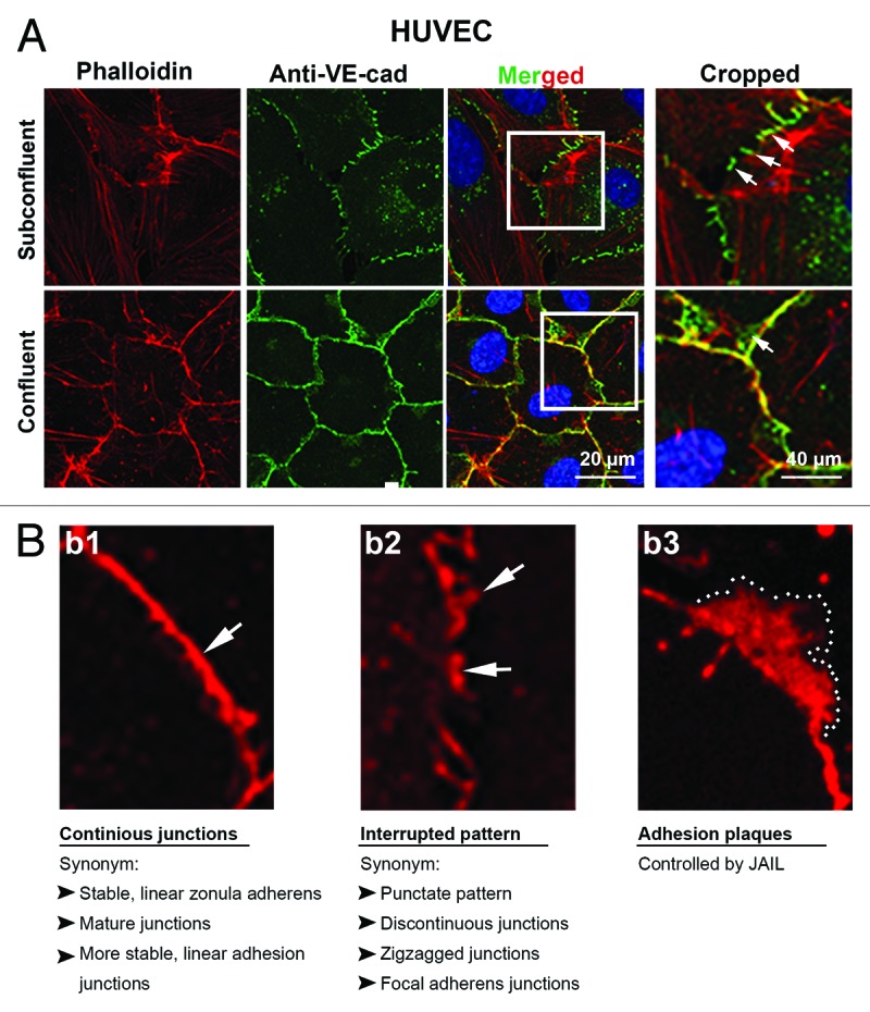
Figure 1. Distribution of VE-cadherin and actin at cell junctions of subconfluent and confluent endothelial cells in culture. Subconfluent and confluent human umbilical vein endothelial cells (HUVEC) cultures were labeled with (a) anti-VE-cadherin (green) and phalloidin-TRITC for filamentous actin (red) or (b1–3) VE-cadherin alone (red). (a) VE-cadherin appears interrupted in the subconfluent cultures (arrows) with large cells and extended perimeter, while cells of confluent cultures are small and polygonal and preferentially exhibits a continuous VE cadherin distribution (arrow). Boxes in the merged images indicated the cropped area as indicated. Actin filaments are incomplete co-localized with the VE-cadherin/catenin complex in both subconfluent and confluent cultures. (b1–3) Higher magnification of HUVEC cultures labeled with anti-VE-cadherin (red). (b1) VE-cadherin appears in confluent cultures in a continuous VE cadherin patterning (arrow) while (b2) in subconfluent cultures, VE-cadherin preferentially appears as an interrupted patterning by which VE-cadherin cluster display different sizes (arrows). (b3) A VE-cadherin adhesion plaques, which is a result of JAIL formation is indicated (dotted line).