
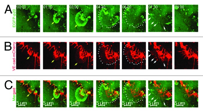
Figure 3. Time-lapse series of ARP2/3 complex-mediated JAIL formation and VE-cadherin dynamics in subconfluent endothelial cell cultures expressing both the fusion protein EGFP-p20 (green) and VE-cadherin-mCherry (red) at high magnification. (A) JAIL developed (yellow arrows) to its maximal extension within 4–5 min (green, dotted lines). JAIL developed close to and between interruptions of VE-cadherin-m-Cherry clusters and caused new VE-cadherin adhesion plaques (B, yellow arrows and dotted lines). (B) VE-cadherin-mCherry plaques (dotted lines) increasingly cluster (white arrows) during JAIL retraction and assemble at cell junctions. This mechanisms change the VE-cadherin pattern, and thus, contributes to VE-cadherin dynamics. Taken from Taha et al., 2014, MBoC.