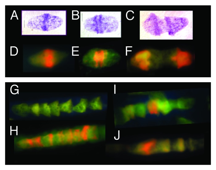
Figure 1. Metakaryotic nuclei undergoing two forms of symmetrical amitoses within tubular syncytia of fetal spinal cord ganglia (9 wks). (A, B, and C) Three “kissing bell” amitoses with DNA stained Feulgen reagent (purple). The order shown represents the stages of (A) formation of two pairs of condensed DNA rings at the rim of the bell mouth that (B) separate as total DNA increases and (C) proceeds toward complete separation when an exact doubling of DNA is observed.3 (D, E, and F) Three “kissing bell” amitoses stained with acridine orange after treatment with RNase. The order shown represents the stages of (D) appearance of bright orange fluorescence co-localized with the condensed DNA rings of Feulgen stained condensed DNA as in (A), (E) separation of two orange rings at the bell mouths as total DNA increases as in (BandF) progress toward complete separation as in (C). (G, H, I, and J) Four lower magnification images of syncytia stained with acridine orange after RNase treatment. (G) A section of a syncytium with well-separated bell shaped nuclei showing the common “stacked cup” relationship of metakaryotic nuclei in tubular syncytia when no amitoses are evident. Well-separated bell shaped nuclei stained with acridine orange emit green fluorescence with scant amount of orange fluorescence. (H) A tubular syncytial section with bell shaped nuclei displaying the closely stacked nuclei indicative of synchronous amitoses and strong orange fluorescence associated in this example with the rims of the bell mouths. (I and J) Two syncytial sections displaying the “stacking cup” form of amitoses with varying degrees of separation between nuclei and the intensity and distribution of orange fluorescence among nuclei within the syncytia. Images by EVG.
