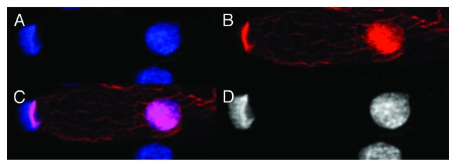
Figure 5. Images of dsDNA (DAPI, blue) and dsRNA/DNA (TRITC-Ab, red) stain in mononuclear metakaryotic cell undergoing asymmetrical amitosis in a colonic adenocarcinoma (M, 68 y). (A) DAPI fluorescence (blue). (B) TRITC-Ab fluorescence (red). (C) Merged images of (AandB) showing nuclei labeled simultaneously with DAPI and TRITC-Ab. (D) Achromatic image of (A). Image is interpreted as an asymmetrical amitosis in which both parent bell shaped nucleus and daughter spherical nucleus have reconverted a large fraction of the dsRNA/DNA intermediate (red) to the dsDNA form (blue). Appearance of red fragments or striations of dsRNA/DNA antibody labeling in cytoplasmic volume between nuclei is occasionally observed as shown here in tumors and in the HT-29 cell line. Image by Koledova VV.
