
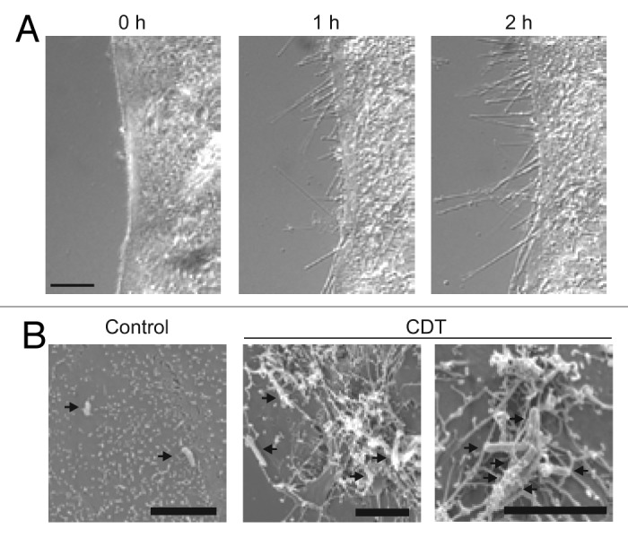
Figure 4. Microscopic pictures of CDT-induced protrusions. (A) The microscopic pictures show protrusion formation after treatment of human carcinoma Caco-2 cells after CDT treatment. (B) Electron microscopic studies show clostridia embedded in the meshwork of protrusions (pictures are modified from Schwan et al.75).