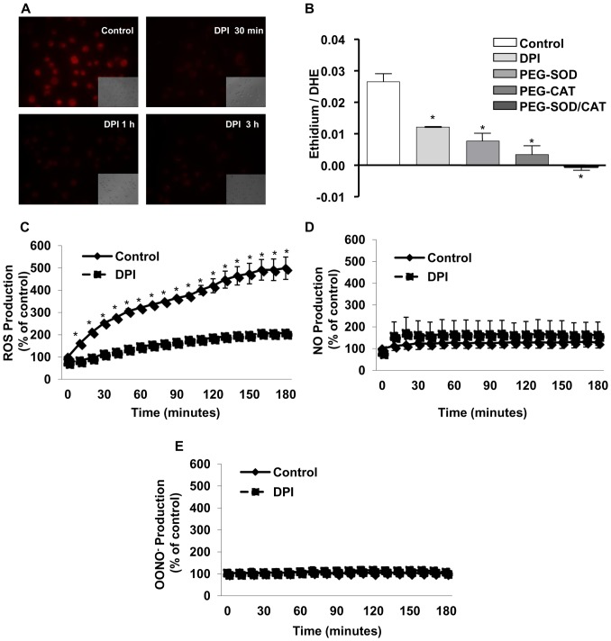
Figure 1. Inhibition of NADPH oxidase activity abolishes intracellular ROS generation on melanoma cells MV3.
(A) After adhesion, melanoma cells were incubated with or without DPI (10 µM) for different times (0.5–3 h) and ROS generation was evaluated by dihydrorhodamine-123 (DHR) assay followed by fluorescence microscopy analysis. (B) MV3 cells were incubated for 1 h with DPI (10 µM), PEG-SOD (25 U/mL), PEG-CAT (200 U/mL), or PEG-CAT and PEG-SOD (200 U/mL, 25 U/mL, respectively). Cellular ROS production was measured by intracellular oxidation of DHE to ethidium assessed by HPLC. (C–E) MV3 cells were incubated with or without DPI (10 µM). Intracellular ROS production was measured by intracellular oxidation of CM-H2DCFDA (C), DAF-AM (D) or HPF (E), as described in Material and Methods. Data are expressed as mean ± SD of three independent experiments. * p<0.05 vs. control;

