
Figure 3. Characterization of ASC and vascular graft creation within the rabbit model.
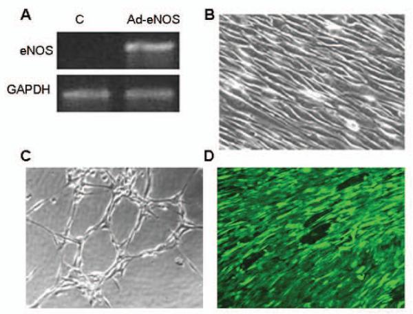
(A) RTPCR analysis of eNOS mRNA expression rabbit ASC differentiated towards and EC lineage before (control, C) and after (Ad-eNOS) eNOS transfection. (B) Phase contrast photomicrograph of rabbit ASC differentiated towards an EC lineage after application of shear stress (1.5 dyne/cm2 at 0.2 Pa, 48h) demonstrating alignment of the cells in the direction of shear. (C) Phase contrast photomicrograph of EC-differentiated rabbit ASC following seeding onto Matrigel demonstrating cord formation indicative of angiogenic potential. (D) Laser confocal micrograph of the luminal surface of a vascular scaffold seeded with differentiated and transfected autologous rabbit ASC and flow conditioned for 5d demonstrates the adherence and alignment of the seeded cells (Cell Tracker Green).