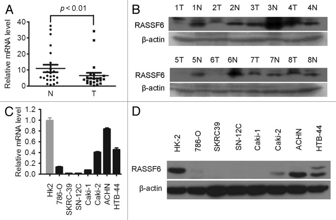
Figure 1. Downregulation of RASSF6 expression in ccRCC tissues and cell lines. (A) RASSF6 mRNA expression levels in 20 matched primary ccRCC tissues (T) and adjacent noncancerous tissues (N) were determined by qPCR assays. GAPDH and 18S were used as reference genes. P < 0.01, paired t test. (B) Western blotting analysis of RASSF6 protein levels in another randomly selected 8 pairs of matched ccRCC tissues (T) and adjacent noncancerous tissues (N). (C and D) qPCR (C) and western blotting (D) analysis of RASSF6 expression in ccRCC cell lines and HK-2 immortalized renal proximal epithelial tubular cells.

An official website of the United States government
Here's how you know
Official websites use .gov
A
.gov website belongs to an official
government organization in the United States.
Secure .gov websites use HTTPS
A lock (
) or https:// means you've safely
connected to the .gov website. Share sensitive
information only on official, secure websites.
