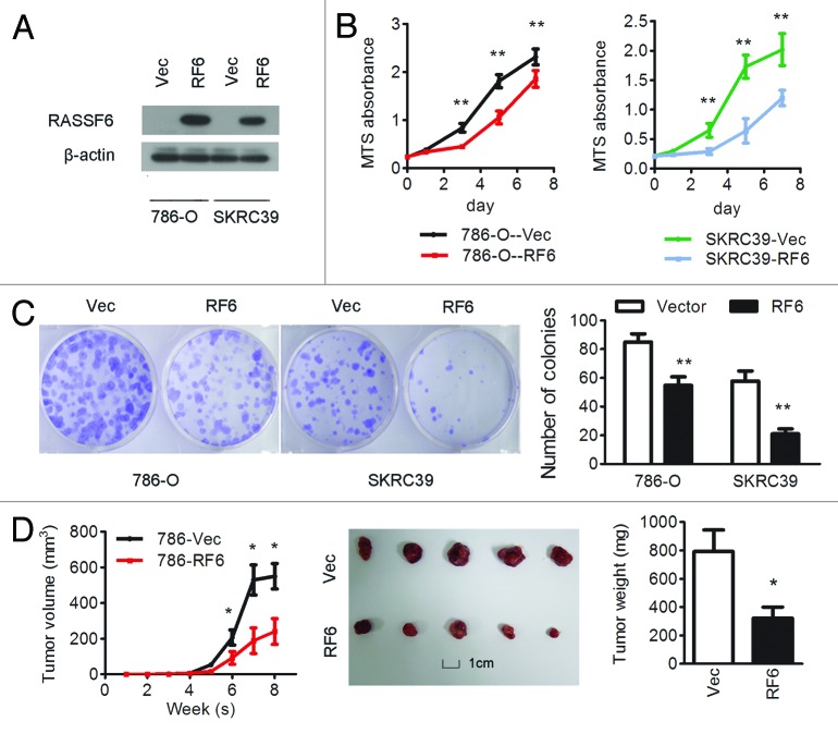
Figure 2. Overexpression of RASSF6 inhibits the proliferation of ccRCC cells in vitro and in vivo. (A–C) 786-O and SKRC39 cells stably overexpressing RASSF6 (RF6) or transfected with empty vector (Vec) were analyzed as follows. (A) RASSF6 protein expression levels were determined by western blot analysis; β-actin was used as a loading control. (B) Cell proliferation was determined by the MTS assay; *P < 0.05, **P < 0.01, Student t test. (C) Colony formation ability; representative micrographs (left) and quantification (right) of crystal violet-stained cells from 3 independent experiments; *P < 0.05, **P < 0.01, Student t test. (D) Control or RASSF6-overexpressing 786-O cells were inoculated subcutaneously into nude mice (n = 5/group). Tumor volumes were measured (left) and weighed (right) on the last day of the experiment. Representative images of isolated tumors (middle) are presented; *P < 0.05, Student t test; scale bar in picture: 1 cm.

An official website of the United States government
Here's how you know
Official websites use .gov
A
.gov website belongs to an official
government organization in the United States.
Secure .gov websites use HTTPS
A lock (
) or https:// means you've safely
connected to the .gov website. Share sensitive
information only on official, secure websites.
