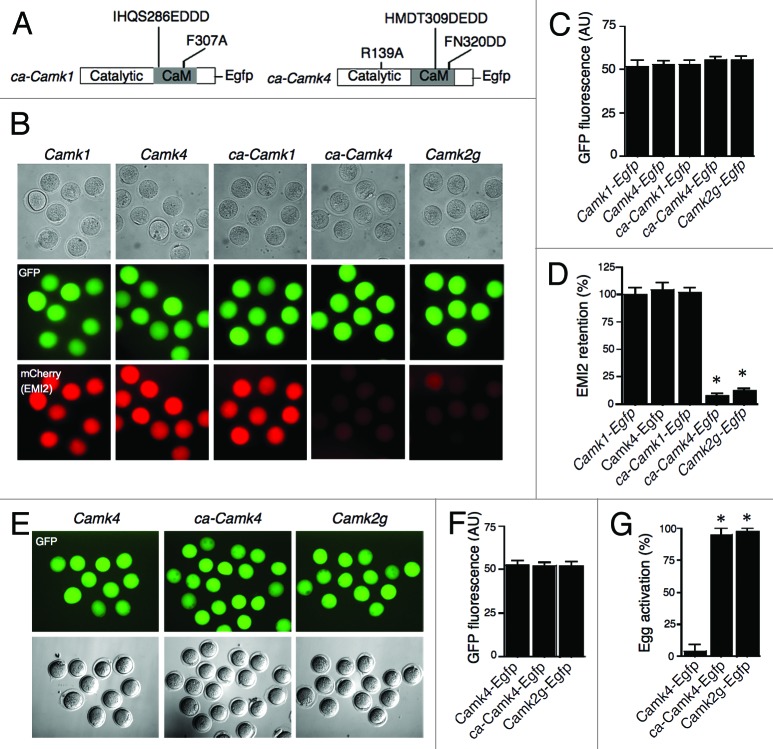
Figure 4. Expression of ca-CaMKIV in CaMKIIγ−/− eggs triggers degradation of exogenous EMI2 and cell cycle resumption. (A) Schematic representation of the mutation strategy to produce constitutively active forms of CaMKI (ca-CaMKI) and IV (ca-CaMKIV). (B) Representative images for the assessment of EMI2 stability in different groups based on mCherry signal. Experimental procedures were the same as described in Figure 2. (C) Quantitative adjustment of expression of different constructs by GFP signal. (D) Quantification of the amount of EMI2 retained in Camk2g−/− eggs. Camk1 group was used as a control, since the experiments depicted in Figure 2 had established that exogenous EMI2 remains essentially intact in the presence of CaMKI. The data are expressed as the amount of EMI2 in Camk4, ca-Camk1, ca-Camk4, and Camk2g groups relative to the amount in Camk1 group. (E–G) Activation of Camk2g−/− eggs expressing different CaMK constructs. Experimental procedures were the same as described in Figure 1. *P < 0.0001 vs. KO eggs injected with Camk1 cRNA. (E) The expression of Camk4, ca-Camk4, and Camk2g was adjusted to similar level by the intensity of GFP signal. Activation of eggs was assessed by PN formation 8 h after the onset of SrCl2 treatment. Magnification: 200×. (F) Quantitative adjustment of expression of different constructs by GFP signal. The data are expressed as the mean ± SEM (G) Egg activation, as measured by pronuclear formation, in the different groups 8 h after exposure to 10 mM SrCl2. The data are expressed as the mean ± SEM. Statistical analysis was performed using ANOVA. *P < 0.0001.

An official website of the United States government
Here's how you know
Official websites use .gov
A
.gov website belongs to an official
government organization in the United States.
Secure .gov websites use HTTPS
A lock (
) or https:// means you've safely
connected to the .gov website. Share sensitive
information only on official, secure websites.
