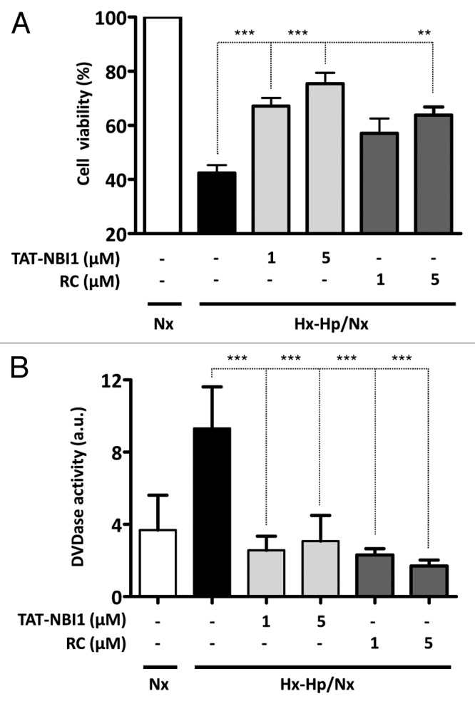
Figure 1. CDK inhibitors increase cell viability in a model of ischemia/reperfusion-induced cell death in renal tubular cells. LLC–PK1 cells were either maintained in Nx (white bars) or subjected to Hx–Hp conditions (1.5% O2; 18% CO2). After 24 h, cells were treated (gray bars) or not (black bars) with TAT-NBI1 (1 and 5 µM) or roscovitine (1 and 5 µM) and were maintained under Nx conditions (21% O2; 5% CO2) for 24 h. (A). Cell survival was measured by the trypan blue exclusion assay. (B) Caspase-3/7 activity was measured under the conditions described above. In all cases, data are expressed as the mean ± SE (n > 3). (***P < 0,0001; **P < 0,01).
