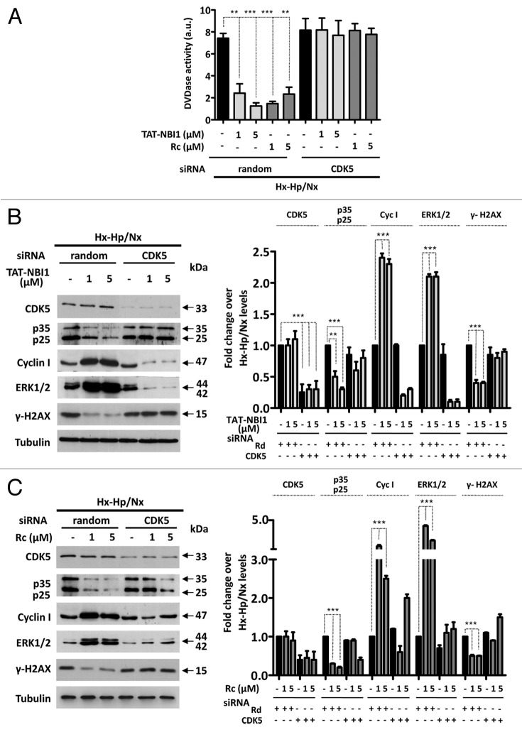
Figure 3. CDK5 is not involved in apoptosis induction, but is required to engage the TAT-NBI1 treatment-induced recovery program. LLC–PK1 cells were untreated or transfected with a control (random) or a CDK5-specific siRNA. After 24 h of silencing, cells were treated as indicated in Figure 1. (A) The caspase-3/7 activity of the cytosolic extracts from treated cells. Error bars represent the mean of 3 experiments ± sd. The expression of CDK5, p35/p25, cyclin I, and the phosphorylation levels of ERK1/2 and H2A.x were analyzed by western blot in the cells treated either with TAT-NBI1 (B) or roscovitine (C). The densitrometric analysis of the western blots from 3 independent experiments are shown in the right-hand panels.

An official website of the United States government
Here's how you know
Official websites use .gov
A
.gov website belongs to an official
government organization in the United States.
Secure .gov websites use HTTPS
A lock (
) or https:// means you've safely
connected to the .gov website. Share sensitive
information only on official, secure websites.
