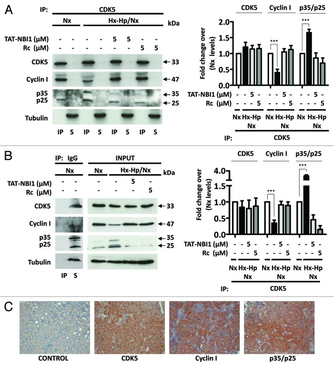
Figure 7. CDK5 inhibitors favored the formation of CDK5/cyclin I complexes to promote cellular recovery mechanisms. Cells were treated as described in Figure 1. (A) Immunoprecipitation (IP) of the CDK5 protein in the LLC–PK1 cell lysates probed with the CDK5, cyclin I and p35/p25 antibodies. S, supernatant. (B). A non-relevant immunoglobulin (IgG) was used as the control for non-specific immunoprecipitation. Control IP and total cell lysates (inputs) were immunoblotted with the CDK5, cyclin I, and p35/p25 antibodies. A densitrometric analysis of the western blots from 3 independent experiments are shown in the right panels. (C) Immunohistochemistry analysis of rat kidneys showing morphological hematoxylin and CDK5, cyclin I and p35/p25 staining of the tubular renal cells.

An official website of the United States government
Here's how you know
Official websites use .gov
A
.gov website belongs to an official
government organization in the United States.
Secure .gov websites use HTTPS
A lock (
) or https:// means you've safely
connected to the .gov website. Share sensitive
information only on official, secure websites.
