Figure 1.
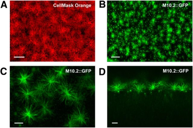
M10.2::GFP enrichment in VSN microvilli. A, Confocal imaging of microvilli covering the VNE surface. Microvilli were visualized in red using the plasma membrane dye CellMask Orange in an ex vivo whole-mount VNO preparation of a WT mouse (en face). Scale bar, 10 μm. B, Confocal imaging of M10.2::GFP fluorescence in whole mount. En face view of the surface of VNE reveals a homogeneous distribution of M10.2::GFP VSNs. Individual microvilli emanating from a given dendritic knob are clearly visible. A z-stack of 60 images was taken (Δz = 0.7904 μm) and a maximum z-projection was produced. Scale bar, 20 μm. C, Single confocal image, en face view, showing M10.2::GFP+ microvilli at higher resolution. Scale bar, 5 μm. D, Single confocal image, side view. The M10.2::GFP fusion protein is strongly enriched in the microvilli. Scale bar, 5 μm.

