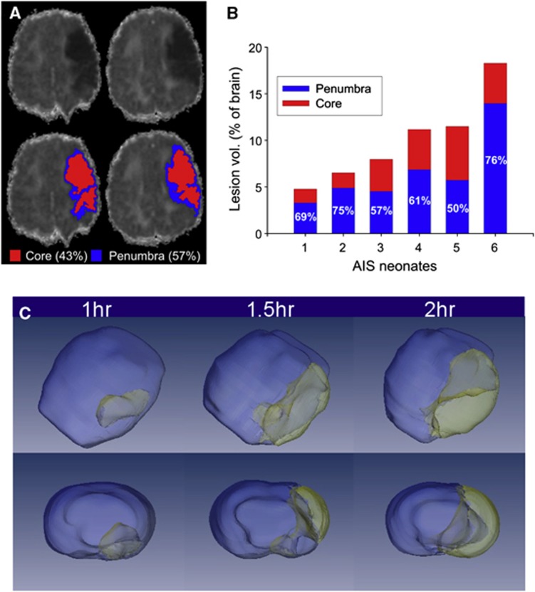
Figure 1.
Magnetic resonance imaging (MRI)-based delineation of the ischemic core and penumbra. (A, B) Diffusion-weighted MRI (DWI) in human term babies 3 to 5 days after arterial ischemic stroke delineates the presence of penumbra (in red). Core and penumbra can be determined using a computational analysis method (Hierarchical Region Splitting) based on apparent diffusion coefficient threshold values as described in Ashwal et al.79 The axis in (B) denotes data from six individual term neonates showing total lesion, core, and penumbral volumes. Note the wide variability in volumes that can occur in neonatal arterial ischemic stroke (AIS). (C) An example of injury volumes in a rat pup model of AIS. The 3D volume of ‘tissue at risk' corresponds to the increasing duration of transient middle cerebral artery occlusion in the postnatal day 10 (P10) rats. Note the increasing size of the ischemic core (yellow) and penumbra (gray) in the hemisphere ipsilateral to the occlusion (courtesy of Stephen Ashwal and Andy Abenous, Department of Pediatrics, Lomo Linda University). These types of translational studies may provide better data regarding injury severity that could be applied to future clinical trials.

