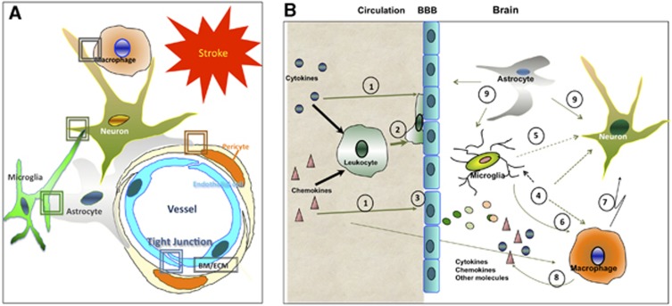
Figure 4.
A schematic diagram that shows major differences in the response of the blood–brain barrier to acute stroke between adults and neonates. (A) Under normal conditions the peripheral and local brain compartments are separated by fine-tuned cell–cell interactions at the neurovascular interface. After stroke, direct damage to several cell types, in concert with activation of immune cells and altered cell–cell communications, adversely affect functional integrity of the blood–brain barrier (BBB). Boxes point to known differences in processes in cerebral cortical regions affected by transient middle cerebral artery occlusion (tMCAO) in adult and neonatal rodents. BM/ECM, basal membrane/extracellular matrix. (B) Events at the neurovascular interface after tMCAO in neonatal rats: 1—peripheral cytokines and chemokines activate leukocytes and prime endothelium; 2—extravasation of neutrophils and monocytes is low, contributing to functional preservation of the BBB. Note that in adult rats after tMCAO leukocytes extravasate into ischemic-reperfused brain regions through distorted BBB; 3—BBB leakage is low compared with major BBB leakage and associated entrance of variety of molecules from peripheral circulation entering the injured adult brain; 4—inflammatory mediators that either enter the brain or are produced by local cells activate resident brain cells, including further activation of microglia, macrophages, endothelial cells, and astrocytes; 5—microglial cells signal to neurons; 6—processes shown as 1 to 3 activate microglial cells, prompting morphologic and functional transformation of these cells to brain macrophages. Peripheral monocytes that enter the brain gradually differentiate to become brain macrophages, but activated microglial cells are the predominant population of brain macrophages after acute injury; 7—activated microglia/macrophages directly signal to neurons; 8—microglia/macrophages produce various cytokines and chemokines; and 9—astrocytes directly communicate with neurons and cells of the neurovascular units. 4 to 9—compared with injury in the adult brain, cell–cell communications differ in the newborn in at least several respects, as described in the text.

