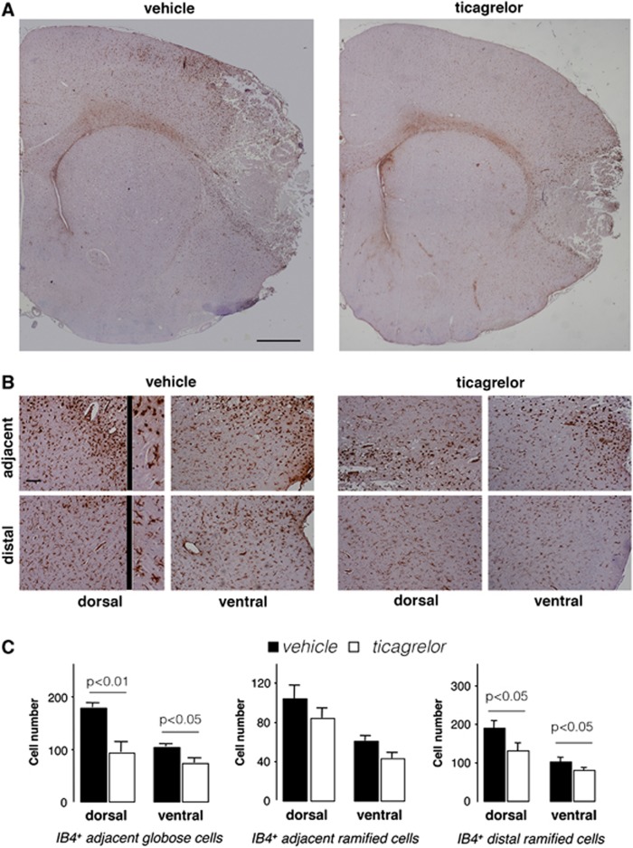
Figure 6.
Effect of ticagrelor on IB-4+ cells in middle cerebral artery occlusion (MCAo) rats. (A) Representative images of IB-4 immunohistochemistry in ischemic brain slices from vehicle- and ticagrelor-treated MCAo rats. Scale bar=1 mm. (B) High magnification of adjacent and distal fields of vehicle and drug-treated ischemic animals. Adjacent fields display both globose and ramified IB-4+ cells (see inset at × 4 magnification), whereas distal fields display only ramified cells (see inset at × 4 magnification). Scale bar=300 μm. (C) Histograms showing the quantitative analysis of the number of IB-4+ globose and ramified cells in adjacent regions and of the number of IB-4+ ramified cells in distal regions in vehicle- and ticagrelor-treated animals. The data represent the mean±s.e.m. of six different rats/group.

