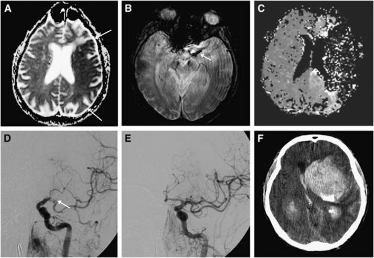
Figure 2.
Recanalizing into an infarcted area. A 76-year-old man with no vascular risk factors except hyperlipidemia, presented with right-sided weakness, aphasia and eye deviation to the left. National Institutes of Health Stroke Scale (NIHSS) was 21. Magnetic resonance imaging (MRI) was performed only 33 minutes after symptom onset. Despite the fast evaluation, MRI showed a large infarct in the left middle cerebral artery (MCA) territory on the apparent diffusion coefficient (ADC) image (A). A clot was found in the left MCA (B). Time-to-peak map showed artifactual zero signal intensity mapping of the left MCA territory due to severe contrast arrival delay (C). Since the patient was without risk factors and in a good state of health, he was taken for intraarterial therapy (IAT) despite the large infarct. On digital subtraction angiography (DSA), the clot was initially found where the internal carotid artery (ICA) divides into the MCA and the anterior cerebral artery (T-occlusion, not shown). After initial removal, DSA now shows the clot in the left MCA (D). Recanalization (E) was achieved using a stent retriever, clot aspiration, and delivery of 4 mg of intraarterial tissue plasminogen activator (tPA) and the patient was taking to the Intensive Care Unit. Minutes after, the patient lost consciousness. An acute computer tomography (CT) scan (F) showed a large intracerebral hemorrhage. The patient died a few hours later.

