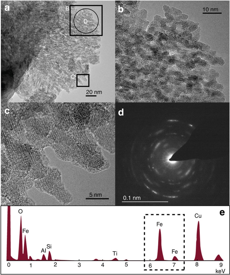
Figure 3. Photomicrographs of LG subglacial suspended sediment.
Nanoparticulate ferrihyrite ~\n5–10 nm in diameter has been identified. Images (b) and (c) are enlargements of (a), as indicated. The diffraction signal (d) shows some crystalline structure owing to possible impact of nano-clay particles, and potentially nano-hematite, but also the characteristic diffuse ferrihydrite rings are identifiable. EDS (e) analysis of the area further confirms Fe-dominated material.

