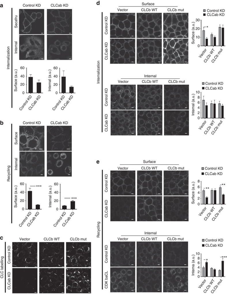
Figure 5. CLC depletion decreases recycling of inactive β1-integrin.
(a) siRNA-treated HeLa cells were allowed to internalize antibody specific for inactive β1-integrin for 30 min at 37 °C. Cells were fixed and stained with Alexafluor-conjugated secondary antibody to visualize surface integrins. For analysis of internal integrins, surface antibody was stripped by acid wash before fixation, followed by permeabilization. Quantification of surface (bottom left) and internal (bottom right) fluorescent labelling of inactive β1-integrin antibody following internalization, which are presented as raw mean fluorescent intensities (mean±s.e.m.; n=3; P=not significant, Student’s t-test). (b) Cells were treated as in a, followed by surface antibody stripping by acid washing after the 30 min internalization period. Cells were then placed at 37 °C for 30 min to chase integrins back to the cell surface. Cells were fixed and processed as in a. Quantification of surface (bottom left) and internal (bottom right) fluorescent labelling of inactive β1-integrin antibody following recycling (mean±s.e.m.; n=3; ***P<0.005, Student’s t-test). (c) HeLa clones expressing the vector, siRNA-resistant WT CLCb or siRNA-resistant mutant (mut) CLCb were treated with control or CLCab-targeting siRNA and immunostained for CLC. (d) Internalization of inactive β1-integrin antibody was assessed as in a in siRNA-treated HeLa clones that expressed the vector, siRNA-resistant WT CLCb or siRNA-resistant mutant CLCb. Quantification of surface (top right) and internal (bottom right) fluorescent labelling of inactive β1-integrin antibody following internalization (mean±s.e.m.; n=3; *P<0.05, two-way analysis of variance (ANOVA) followed by Bonferroni post hoc test). (e) Recycling of inactive β1-integrin antibody was assessed as in b in siRNA-treated HeLa clones that stably expressed the vector, siRNA-resistant WT CLCb or siRNA-resistant CLCb. Quantification of surface (top right) and internal (bottom right) fluorescent labelling of inactive β1-integrin antibody following recycling (mean±s.e.m.; n=3; *P<0.05, **P<0.01, ***P<0.005, two-way ANOVA followed by Bonferroni post hoc test). Scale bars, 7.5 μm for all panels. a.u., arbitrary unit.

