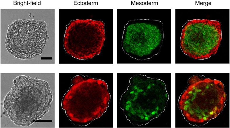
Figure 9. Confocal images of ectoderm and mesoderm germ layers within the same colony.
Representative images of two different colonies with bright field (left column), a distinct Pax6 ectoderm immunofluorescence at the outer layer (2nd column), a distinct Brachyury mesoderm immunofluorescence at the middle layer (3nd column) and merge fluorescence images (right column). Scale bar, 50 μm. From these staining data and those staining data of the colonies in the movies, assuming individual cells from each germ layer have the same volume, we estimate that endoderm, mesoderm and ectoderm layer cells are ~5%, 60% and 35% of the total cells, respectively. These results are representatives of >3 experimental replicates.

