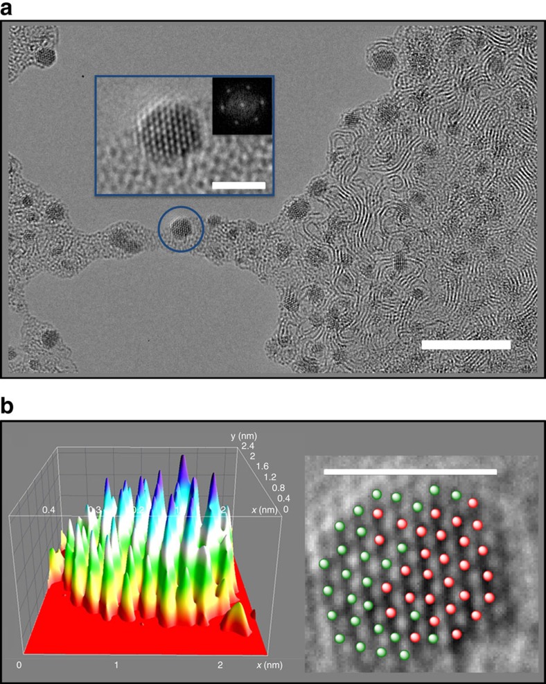
Figure 3. Ru-Os 3D-nanocrystals from RuOsMs micelles.
(a) An array of mixed metal nanocrystals on the graphitic support formed after 60 min of irradiation (scale bar, 10 nm), with enlargement of the blue circled crystal showing atomic resolution (scale bar, 2 nm) and fast Fourier transform analysis of the hexagonal mixed metal crystal. The fast Fourier transform analysis of the hexagonal mixed metal crystal is also shown in the enlargement. (b) 3D projection (left) of the same crystal showing the difference of contrast between Ru, Os and the background (each peak corresponds to an atom, and the height/intensity of the peaks is dependent on the atomic TEM contrast; arbitrary colours), also depicted as 29 red (Os) and 28 green (Ru) balls on the 2D projection (right, scale bar, 2.1 nm). The presence of Ru and Os atoms was confirmed by a combination of scanning-TEM and energy-dispersive X-ray analysis.

