
Figure 5.
hNPC-Mediated Recovery Is Associated with Treg Emergence within the Spinal Cord
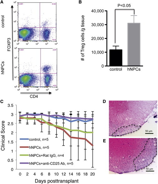
(A–E) Mice were infected intracranially with JMHV and transplanted with either hNPCs or vehicle alone at day 14 post-infection.(A) Examination of Tregs within the spinal cords by flow analysis (CD4+CD25+FOXP3+) revealed an increased frequency of Tregs in hNPC-transplanted animals at day 10 posttransplantation, a time at which animals began to display improved motor skills compared to control mice. A representative flow cytometric dot blot is shown. (B) Quantification of Treg numbers in spinal cords of mice indicated a significant (p < 0.05) increase in the number of Tregs in hNPC-transplanted mice versus controls from 8–10 days posttransplantation. Data are representative of three independent experiments with a minimum of three mice per group; data are presented as average ± SEM. Mann-Whitney t tests were used to determine the p values. (C) hNPC-transplanted mice receiving anti-CD25 antibody did not display recovery in motor skills as compared to hNPC-treated mice treated with control antibody. (D and E) H&E staining of spinal cords of hNPC-treated mice treated with either control antibody (D) or anti-CD25 (E) indicates that depletion of Tregs increases neuroinflammation within white matter tracts (representative images are shown).