Figure 4.
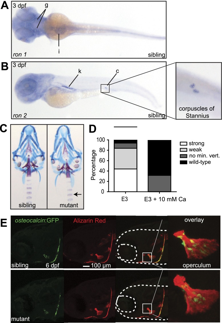
Excess calcium rescues the msp mutant phenotype and restores osteocalcin expression. A, B) Whole-mount in situ hybridization of 3-dpf embryos showing increased expression of ron-1 (A) in the gills (g) and intestine (i) (ventral view) and ron-2 (B) in the kidney (k) and corpuscles of Stannius (c) (lateral view). Inset: higher magnification of the corpuscles of Stannius. C) Alizarin red and Alcian blue staining of 6-dpf wild-type vs. mutant embryos treated with 10 mM calcium in E3 medium. Note the complete phenotypic rescue of mutant individuals, including Alizarin red staining in the vertebrae (arrow). D) Bar graphs represent percentages of rescued mutant embryos in E3 medium compared E3 medium with 10 mM calcium at 6 dpf (average of 3 independent experiments, n=12/experiment). E3 medium with 10 mM calcium significantly rescued the msp mutant phenotype when compared to E3 medium (P<0.001 by chi square test). E) Osteocalcin:GFP+ osteoblasts are present in 6-dpf sibling and mutant embryos treated with 10 mM calcium in E3 medium. Insets: magnification of the operculum, showing the presence of mature osteoblast cells.

