
Fig. 2.
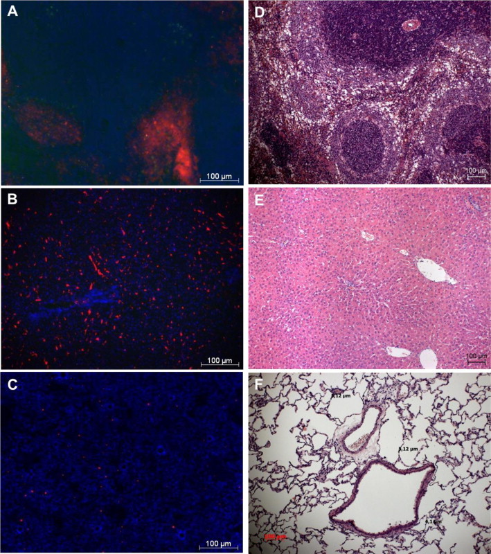
Organ distribution of PFD-filled PLGA microcapsules and effect of capsule infusion on tissue damage. PFD-filled PLGA microcapsules (1.5 µm) were stained with Nile red and were allowed to circulate for about 40 min within the blood circulation, before organs were cryopreserved and frozen. Cryosections of spleen (A), liver (B) and lung (C) were prepared and nuclei were stained with DAPI (blue) for fluorescence microscopic analysis (×200 magnification). For assessment of the effect of capsule infusion on tissue damage, unstained PFD-filled PLGA microcapsules (1.5 µm) were infused for 30 min (20 ml/kg body weight × h). Microscopic assessment (×100 magnification) of spleen (D), liver (E) and lung tissue (F).