
Figure 3.
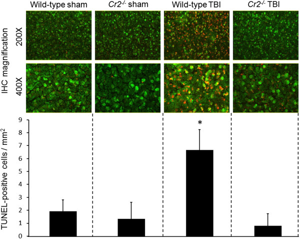
Neuronal cell death is reduced in the injured brain hemispheres of Cr2-/- mice at seven days after traumatic brain injury (TBI). Coronal cryosections of injured left hemispheres of wild-type and Cr2-/- mice at seven days after sham operation or closed head injury were double-stained with a monoclonal anti-NeuN in green fluorescence (FITC) and by TUNEL technique in red fluorescence (TRITC), followed by visualization of cellular signals by fluorescence microscopy. Semi-quantitative analysis of cell-counts revealed a significantly increased number of TUNEL-positive neurons in the injured cortex of wild-type animals, compared to head-injured Cr2-/- mice and sham-operated controls. The TUNEL-positive cells were counted in 15 randomly selected cortical fields of 0.01 mm2 per section. Cell counts are shown as mean values ± SD. *P <0.05 for wild-type TBI compared to Cr2-/- TBI and sham controls. IHC, immunohistochemistry; TBI, traumatic brain injury.