Figure 5.

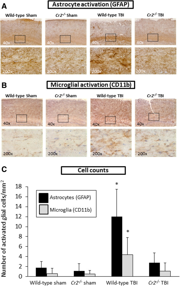
Glial reaction is attenuated in Cr2-/- mice at 24 hours after traumatic brain injury (TBI). Immunohistochemistry of coronal brain cryosections was performed using glial fibrillary acidic protein (GFAP) as a specific marker for astrocytes (A) and CD11b as a cellular marker for microglia (B). The inserts in the upper panel (40 ×) reflect the according selection in the corresponding higher magnification images (200 ×). Cell counts of immunostained sections revealed that signals for GFAP and CD11b were significantly increased in the injured left hemispheres of wild-type mice, compared to head-injured Cr2-/- littermates (C). Activated glial cells were counted in 10 randomly selected cortical fields of 0.01 mm2 per section and per cell marker. Cell counts were performed using QCapturePro7 software (QImaging).and values are shown as mean values ± SD. *P <0.05 for wild-type TBI compared to Cr2-/- TBI and sham controls.
