
Figure 6.
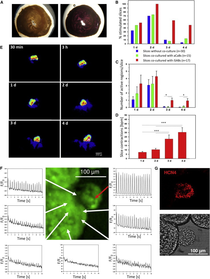
Functionality of iSABs in an Ex Vivo Model of Cultivated Murine Ventricular Slices
(A) Ventricular slices were cultivated for 5 days (left) and MTT staining was used to assess viability (right).
(B) Percentage distribution of iSAB-seeded, aCaB-seeded, and nonseeded slices containing at least one contracting region over time. Spontaneous activity ceased after day 2, whereas activity in iSAB-seeded slices was greatly retained (n ≥ 15).
(C) Average number of active regions per iSAB-seeded, aCaB-seeded, and unseeded slices over time. The data represent three independent experiments and are presented as means ± SEM; n ≥ 15; ∗p < 0.05.
(D) Increase of beating frequencies of iSAB-seeded slices from day 1 until day 4. The data represent three independent experiments and are presented as means ± SEM; n ≥ 16; ∗∗∗p < 0.05. See also Movie S7.
(E) Transmission of calcein dye from an iSAB to the recipient slice over time. Scale bar, 200 μm.
(F) Stimulation of slice regions (white arrows) in the proximity of an iSAB (red arrow) is accompanied by highly synchronized Ca2+ transients.
(G) The identical iSAB/slice coculture as shown in (F) was stained for HCN4 expressing the nodal cell marker in the iSAB. See also Movie S8.