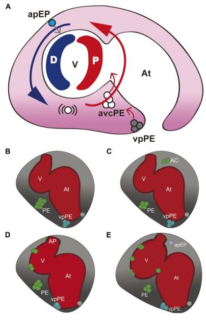
Figure 1. Schematic representation of epicardium formation in the zebrafish.
(A) The model describing the influence of the heartbeat on proepicardium (PE) cluster formation and epicardium morphogenesis. Pericardial fluid flow advects released PE cells around the ventricle until they attach to it. Large blue and red arrows indicate flow direction and flow force (blue, low force; red, high force). Small red arrows indicate the release of cells from the PE clusters; the small gray arrow indicates the transfer of epicardial precursor cells to the myocardium. (B-E) The time frame of events leading to epicardium formation in the zebrafish. (B) At 55 hours postfertilization (hpf), a large PE cluster emerges from the mesothelial wall close to the atrioventricular canal of the forming heart. While two-sided expression of Epi:green fluorescent protein (GFP)-positive cells can be observed before 60 hpf, only the right venous pole PE (vpPE) cluster forms. (C) Over the next 10–12 hours, cells from these clusters are released into the pericardial cavity (gray shading). (D) Advected cells adhere first to the distal ventricle and later to the proximal part. (E) Once attached, epicardial cells proliferate and subsequently flatten. Single cells delaminate from the cranial pericardial mesothelium (arterial pole epicardial precursor pool (apEP)) and are transferred to the ventricular surface. The outflow tract of the heart is covered by pericardial mesothelial cells, which are not derived from the PE clusters. AC, advected cells; apEP, arterial pole epicardial precursor cell; At, atrium; avcPE and PE, atrioventricular canal proepicardial cluster; D, distal; P, proximal; V, ventricle; vpPE, venous pole proepicardial cluster. Images adapted from the original article in Current Biology [35].

