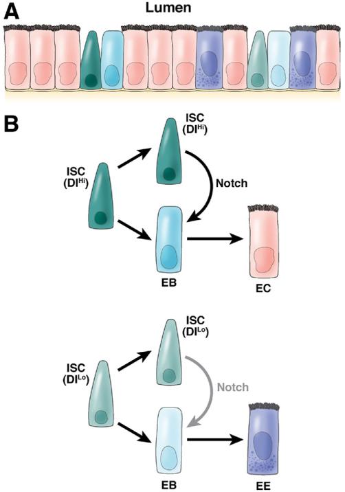
Figure 3. Drosophila posterior midgut and role of Notch in ISC renewal.
(A) The Drosophila posterior midgut is a relatively simple pseudostratified epithelium, with only 2 differentiated cell types: ECs and EE cells. In addition, ISCs and their nonproliferative daughters, EBs, are scattered in between. (B) Two populations of ISCs exist: one with high Delta expression (ISC [DlHi]) and one with no or low Delta expression (ISC [DlLo]). When they divide, both give one daughter ISC and one daughter EB cell. However, the daughter ISC from ISC (DlHi) will activate a high level of Notch signaling in its sister EB cells, which drives its differentiation into EC cells; in contrast, the daughter ISC from ISC (DlLo) elicits a low level of Notch signaling in its sister EB cells, which will differentiate into EE cells. Thus, different levels of Notch activation elicit different cell fates.

