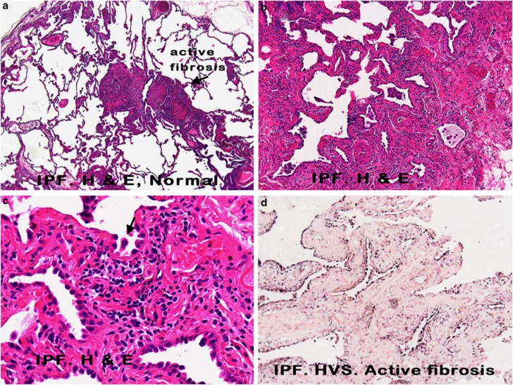
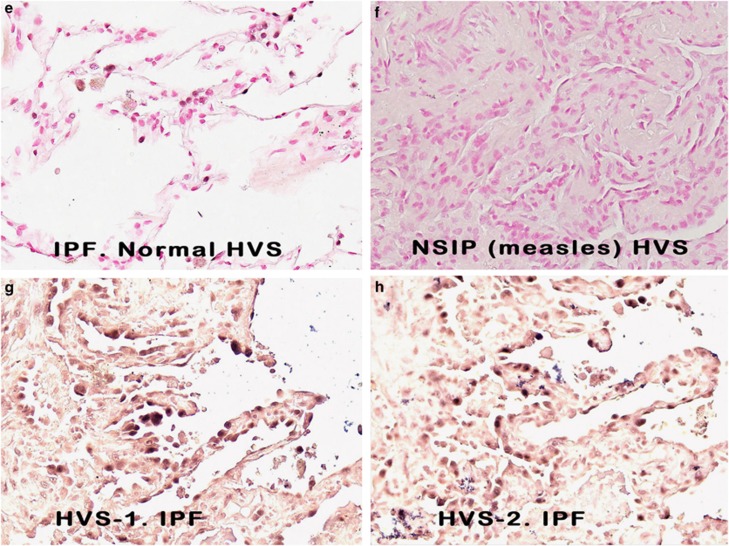
Figure 1.
Histopathological and molecular correlates of idiopathic pulmonary fibrosis. a–c (hematoxylin and eosin) show increased magnifications of idiopathic pulmonary fibrosis in which areas of histologically normal lung is admixed with areas of active fibrosis (a, × 25), marked by serpentine glands lined with epithelia (b, × 50), which show nuclear atypia and multinucleation (c, arrow, × 400). Herpesvirus saimiri DNA was commonly detected in the nuclei of the epithelia of these serpentine glands (d, × 50) and much less evident in the admixed histologically normal lung (e, × 400; blue NBT/BCIP signal with pink counterstain). In comparison, herpesvirus saimiri DNA was not evident in cases of interstitial pneumonitis and fibrosis of known viral etiology (f, × 400 measles infection). Herpesvirus saimiri DNA was evident in the regenerating epithelia in idiopathic pulmonary fibrosis in the same cells when serial sections were probed for herpesvirus saimiri terminal repeat sequence (g, × 400) or the STP ORF 1 gene (h, × 400).


