Figure 2.
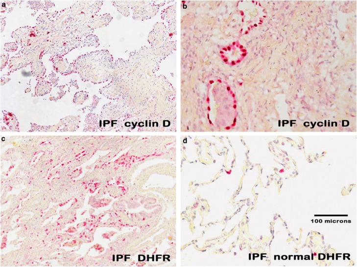
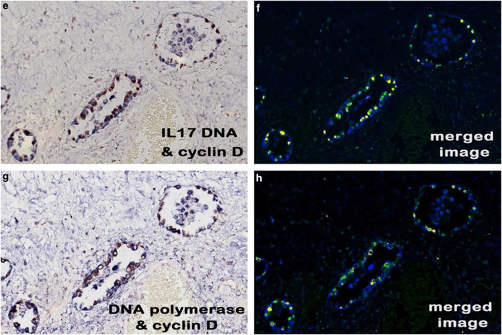
Expression of herpesvirus saimiri-related proteins in idiopathic pulmonary fibrosis. a ( × 100, fast red stain and hematoxylin counterstain) and, at higher magnification, b ( × 400) show that cyclin D1 is highly expressed in the regenerating epithelial cells of idiopathic pulmonary fibrosis. c ( × 100) shows the same pattern of localization for dihydrofolate reductase to the regenerating epithelia in the areas of active fibrosis of idiopathic pulmonary fibrosis and the rarity of the protein in the adjacent normal areas of the same lung tissue (d, × 200). The tissues in e–h (each at × 400) represent photographs of the same cells in serial sections of a case of idiopathic pulmonary fibrosis. The samples were analyzed for either herpesvirus saimiri using an probe specific for the IL-17 DNA sequence (e, f) or the herpesvirus saimiri DNA polymerase gene (g, h). After in situ hybridization, co-expression analysis was performed using immunohistochemistry with either cyclin D (e, f) or dihydrofolate reductase (g, h). In each case, the signal for the DNA was blue and the protein was brown; the Nuance-merged image shows the DNA as fluorescent blue and the protein as fluorescent green, whereas cells with both targets show fluorescent yellow. Note that the same pattern of co-expression is seen in e–h in the merged images.


