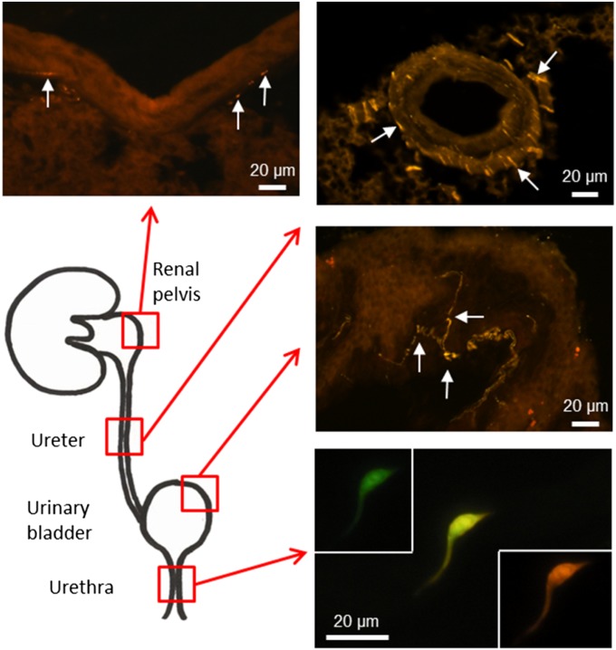
Fig. 1.
Cholinergic eGFP-expressing epithelial cells are restricted to the urethra in the urinary tract. Visualization of GFP was enhanced by application of a chicken anti-eGFP antibody, followed by Cy3-conjugated anti-chicken Ig. GFP and Cy3 channels are shown separately and also in the merged image for the slender epithelia cell in the urethra. In all other parts of the urinary system, cholinergic nerve fibers (arrows) were visualized by this procedure, but no ChAT-eGFP–expressing epithelial cells were seen.

