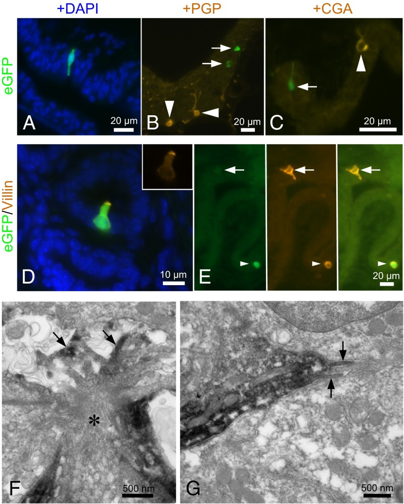
Fig. 2.
A cholinergic epithelial cell of the urethra is a brush cell, not a neuroendocrine cell. (A) Male penile urethra. A slender ChAT-eGFP cell extends through the epithelial layer. Nuclei are counterstained with DAPI. (B) Female urethra. ChAT-eGFP cells (arrows) are distinct from protein gene product 9.5 (PGP)-immunoreactive neuroendocrine cells (arrowheads). (C) Male urethra. The ChAT-eGFP cell (arrow) does not express the neuroendocrine cell marker chromogranin A (CGA). The arrowhead indicates a CGA+ neuroendocrine cell. (D) Male urethra, colliculus seminalis. Merged image showing eGFP (green), villin immunoreactivity (orange), and cell nuclei (DAPI; blue). The flask-shaped ChAT-eGFP cell is immunoreactive for the brush cell marker villin. (Inset) Villin only, showing the concentration of this microvillous protein at the apical cell pole. (E) Male urethra. Another villin+ brush cell is not ChAT+ (arrows). Arrowheads indicate double-positive cells. (F) Female urethra, pre-embedding ultrastructural eGFP immunolabeling. The apical region of the eGFP-immunoreactive cell (asterisk) exhibits characteristic brush cell features, with numerous filaments, tubulovesicular structures, and microvilli (arrows) projecting into the lumen. In this particular case, the microvilli extend into an intercellular cavity in the upper layer of the epithelium that had no visible connection to the urethral lumen. (G) Same cell as depicted in F exhibiting lateral microvilli (arrows), another brush cell feature.

