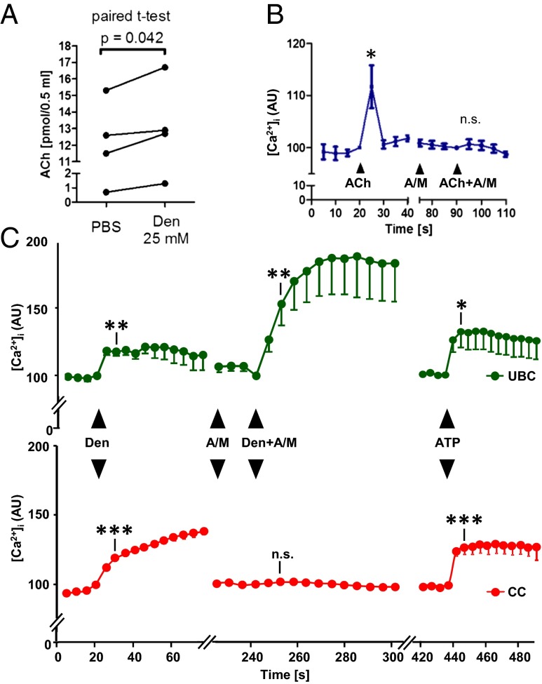
Fig. 5.
Urethral brush cells release acetylcholine. (A) Acetylcholine (Ach) content in the supernatant (0.5 mL) of isolated urethral cells after a 5-min exposure to PBS (control) or denatonium. Isolates from one urethra are treated as paired data. (B) Calcium recordings from eight eGFP− urethral cells located on coverslips that did not contain eGFP+ cells. In the continuous presence of the acetylcholine esterase inhibitor physostigmine (5 µM), acetylcholine (25 µM) evokes an increase in [Ca2+]i that is sensitive to a mixture (A+M) of muscarinic (2 µM atropine) and nicotinic (20 µM mecamylamine) acetylcholine receptor blockers. (C) Parallel [Ca2+]i recordings from eGFP+ (n = 13, Upper) and eGFP− (n = 74; Lower) isolated urethral cells located in their vicinity (same field of view during confocal laser scanning recording) on the coverslip. Both cell types respond to denatonium (Den; 25 mM) with a [Ca2+]i increase. This responsiveness is lost in eGFP− cells, but persists (even at enhanced levels; P = 0.04, paired t test) in eGFP+ cells after pretreatment with cholinergic blockers (A+M; A), demonstrating denatonium-evoked cholinergic signaling from eGFP+ to eGFP− cells. Response to the nongustatory stimulus ATP demonstrates the viability of cells at the end of the experiment. n.s., nonsignificant. *P < 0.05; **P < 0.01; ***P < 0.001, paired t test compared with value immediately before substance application.

