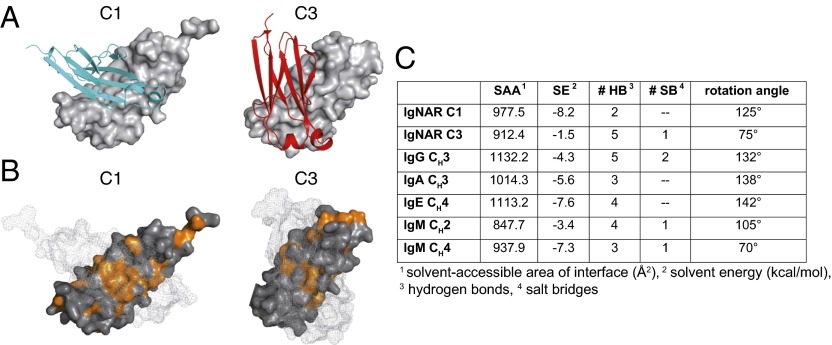
Fig. 2.
Characterization of the IgNAR C1 and C3 dimerization interfaces. (A) Ribbon and surface diagram of the IgNAR C1 and C3 dimers. The two subunits are colored in cyan and gray (C1) or red and gray (C3), respectively. (B) Hydrophobic residues within the C1 and C3 dimerization interfaces are shown in orange. One monomer is in surface representation; its counterpart is shown as a mesh surface. (C) Comparison of the dimerization interfaces of IgNAR C1 and C3 and different human and murine dimeric domains, as determined by the PISA server (48) (Protein Data Bank ID codes IgG CH3, 3HKF; IgA CH3, 1OW0; IgE CH4, 1O0V; IgM CH2, 4JVU; and IgM CH4, 4JVW).

