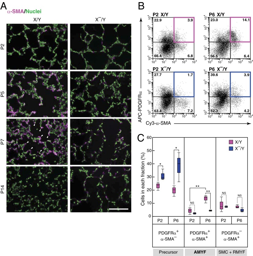
Fig. 2.
Differentiation of AMYFs is blocked during alveolar formation in DA-Raf–deficient mice. (A) AMYFs expressing α-SMA (magenta) were examined by immunohistochemistry in lungs isolated from X/Y and X–/Y mice at various postnatal ages. Arrowheads indicate the tips of alveolar septa. Nuclei are stained with Hoechst dye (green). (Scale bar, 100 μm.) (B) AMYFs expressing PDGFRα and α-SMA were determined by flow cytometric analysis by using the cell suspensions isolated from X/Y (Upper) and X–/Y (Lower) lungs at P2 (Left) and P6 (Right). The x axis and y axis represent Cy3-α-SMA and APC-PDGFRα fluorescent intensities, respectively. The colored boxes show AMYF fractions. (C) Percentages of each quadrant in three independent mice per experimental groups are summarized. The fractions of AMYF precursor, AMYF, and SMC+RMYF contain PDGFRα+/α-SMA– cells, PDGFRα+/α-SMA+ cells, and PDGFRα–/α-SMA+ cells, respectively. **P < 0.01, *P < 0.05; NS, P > 0.05.

