Fig. 3.
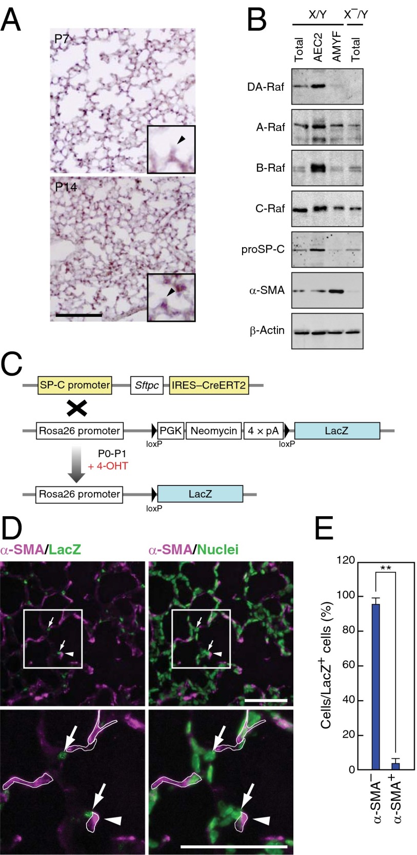
DA-Raf is mainly expressed in AEC2s that are distinct from AMYF precursors. (A) DAraf mRNA was analyzed by in situ hybridization with a probe targeting the 3′ UTR of DAraf mRNA in lung sections from X/Y mice at P7 (Upper) and P14 (Lower). Enlarged images containing alveolar corners and alveolar septa (arrowheads) are shown in the inset of both images. (Scale bar, 100 μm.) (B) Lysates of total lungs, AEC2s, and AMYFs were isolated from X/Y or X–/Y mice at P5 as described in Materials and Methods. These lysates were subjected to Western blotting to analyze the protein levels of DA-Raf and Raf family members. The markers for each fraction were analyzed with anti–proSP-C and anti–α-SMA. (C) Schematic representation of the strategy for lineage-trace of AEC2s. 4-OHT was subcutaneously injected into the pups generated from the crossing SP-C-CreERT2 knock-in mice with ROSA26-LacZ mice at P0 and P1. In the presence of 4-OHT, CreERT2 expressed in AEC2s translocates into the nucleus and mediates excision of the loxP-flanked neomycin cassette, which results in LacZ labeling of AEC2s. (D) Lineage-traced lungs were analyzed by staining for α-SMA and LacZ (Left) at P5. The same slide was stained with Hoechst dye (green) to visualize the nucleus of each cell (Right). Lower panels show higher-magnification images of enclosed squares in Upper panels. The borders of α-SMA+ cells are shown in white lines in Lower panels. Arrows and arrowheads indicate the lineage-labeled cells and alveolar septa, respectively. (Scale bar, 100 μm.) (E) Average percentages of α-SMA+ or α-SMA– cells per lineage-labeled cells were determined in lungs from P5 mice. The values represent means ± SD of different 50 labeled cells from three independent individuals. (**P < 0.01).

