Fig. 5.
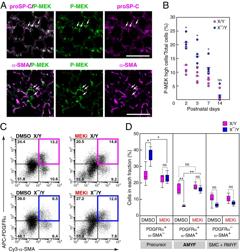
DA-Raf–dependent inactivation of MEK1/2 in AEC2 promotes AMYF differentiation during alveolar formation. (A) Distribution of phosphorylated MEK1/2 (P-MEK) was analyzed in lungs of X/Y mice at P5 by immunohistochemistry. Double-immunofluorescence staining for P-MEK (green) and proSP-C (Upper) or α-SMA (Lower) was performed. Arrows indicate representative cells with high P-MEK levels. (Scale bar, 100 μm.) (B) The percentages of cells containing high levels of P-MEK in lungs of X/Y (pink circles) and X–/Y (blue circles) mice were analyzed at the indicated postnatal ages, as described in Materials and Methods. Circles and lines indicate individual mice and median values, respectively. *P < 0.05; NS, P > 0.05. (C) Representative FACS dot plots of the lung cell suspensions from X/Y (Upper) and X–/Y (Lower) mice treated with DMSO (Left) and MEKi, PD0325901, (Right). The x axis and y axis represent Cy3-α-SMA and APC-PDGFRα fluorescent intensities, respectively. (D) Percentages of each quadrant in four independent mice per experimental groups are summarized. The fractions of AMYF precursor, AMYF, and SMC+RMYF contain PDGFRα+/α-SMA– cells, PDGFRα+/α-SMA+ cells, and PDGFRα–/α-SMA+ cells, respectively. **P < 0.01, *P < 0.05; NS, P > 0.05.

