
Fig. 2.
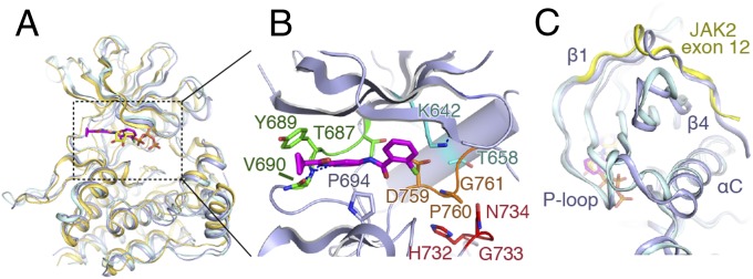
Comparison of the TYK2, JAK1, and JAK2 pseudokinases. (A) Overlay of the TYK2 pseudokinase domain (blue) with the pseudokinase from JAK1 (PDB ID code 4L00; gold) and JAK2 (PDB ID code 4FVQ; cyan). Protein chain is shown as a cartoon model, with ligands shown as sticks (TYK2-bound compound 7012 in magenta, JAK2-bound ATP in yellow). (B) Close-up view of the TYK2 pseudokinase active site. Compound 7012 is shown in magenta, and the positions of canonical kinase features colored [hinge: green; HGN (∼“HRD”) motif: red; DPG (∼“DFG”) motif: orange; αC-helix salt bridge residues: cyan]. (C) View of the exon 12 N-terminal segment of JAK2 compared with TYK2. Protein chains are colored as in A, with the JAK2 exon 12 segment colored yellow.