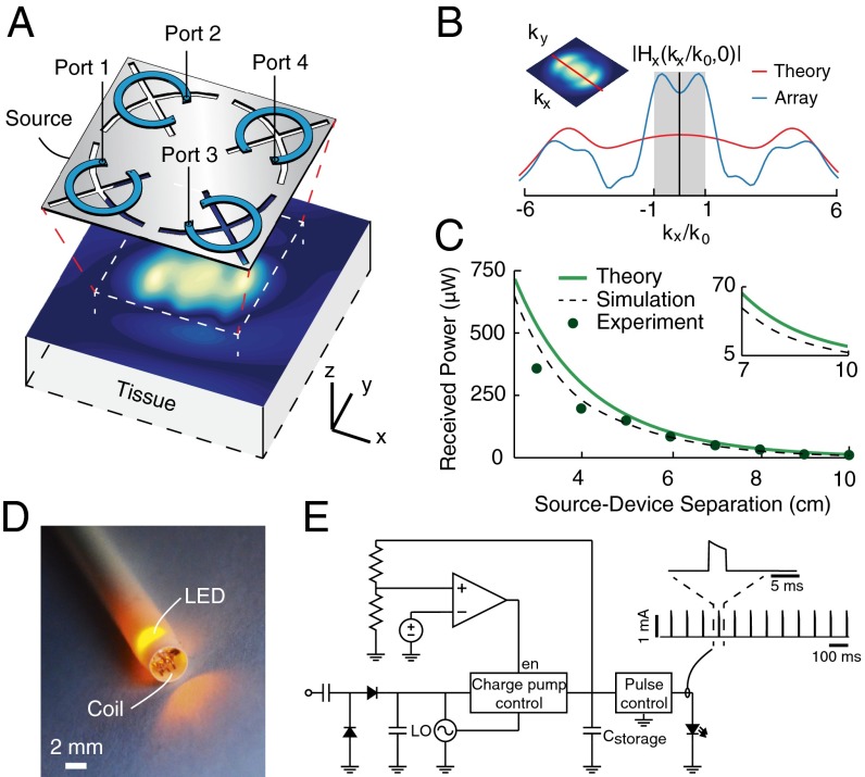
Fig. 2.
Midfield energy transfer with a patterned metal plate. (A) Schematic of the midfield source (dimensions 6 × 6 cm, operating frequency 1.6 GHz) and the magnetic field (time snapshot ) on the skin surface. (B) Corresponding spatial frequency spectrum along the axis compared with the theoretical optimum. (C) Theoretical, numerically simulated, and measured power received by a 2-mm diameter coil as a function of distance when coupling 500 mW into tissue. (D) Photograph of the device inserted in a 10 French (∼3.3 mm) catheter sheath for size comparison. The device is configured as a power detector, containing a 2-mm diameter coil, integrated circuits, and a LED for pulse visualization. (E) Circuit schematic of device with a representative sequence of stimulation pulses generated over 1 s. , charge storage capacitor; en, enable; LO, local oscillator.

