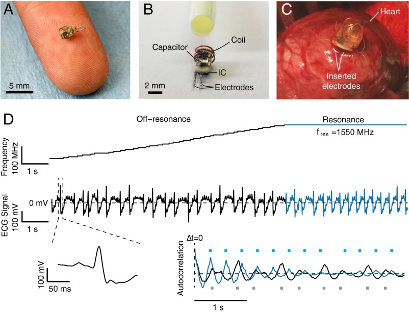
Fig. 5.
Wireless cardiac pacing. (A) Photograph of the wireless electrostimulator on a human finger. The device is 2 mm in diameter. (B) Same device before epoxy encapsulation next to a 10-French (∼3.3 mm) catheter sheath for size comparison. (C) Photograph of the electrostimulator inserted in the lower epicardium of a rabbit via open-chest surgery. After implantation, the chest was closed and a portable, battery-powered source was positioned ∼4.5 cm (1-cm air gap, ∼3.5-cm chest wall) above the device. (D) The operating frequency of the source (Top) adjusted to the estimated resonant frequency of the circuit and maintained for several seconds. Cardiac activity of the rabbit monitored by ECG (Middle), magnified view of a single heartbeat (Bottom Left), and autocorrelation function (Bottom Right) of both the off-resonance (black) and resonant (blue) sections of the ECG signal. Peaks in the autocorrelation function are marked with squares.

