Figure 1.

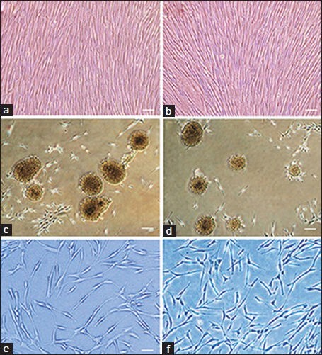
Phase contrast image of (a) hBMSCs and (b) hADSCs Both kinds of cultures were filled with elongated fibroblast-like cells. (c and d) Neurospheres dissociated from the tissue culture dish plastic substrate after 7 days culturing, surrounded by some fibroblast-like cells, almost all of the cells participate in neurosphere formation. (c) Spheres from BM samples culturing; (d) spheres derived from adipose tissue culturing. Differentiated cells derived from (e) hBMSCs and (f) hADSCs 2 weeks after neural induction. We can see bi- and multipolar cells with elongated processes. Scale bars in a and b = 150 μm, c and d = 200 μm and in e and f = 100 μm
