Figure 5.
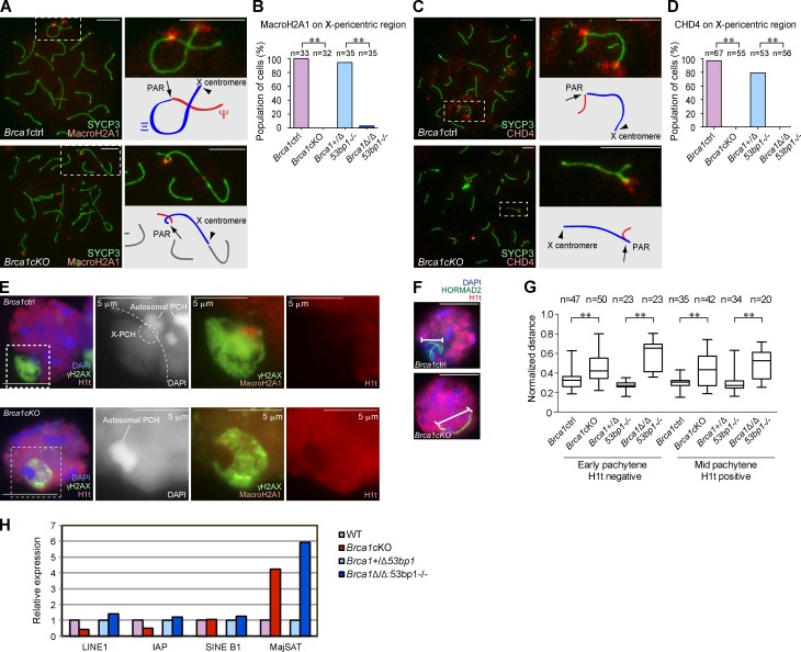
BRCA1 establishes PCH on the X chromosome in male meiosis. (A and C) Immunostaining of meiotic chromosome spreads using anti-SYCP3 and -MacroH2A1 or -CHD4 antibodies. Arrows indicate the PAR. Arrowheads indicate the X centromere. (B and D) Percentage of mid-pachytene spermatocytes with MacroH2A1 or CHD4 on the X-pericentric region. (E) Immunostaining of mid-pachytene spermatocytes using slides that preserve the three-dimensional structure of nuclei. Single Z sections are shown. The regions inside of the dotted squares are shown in the right panels. In the Brca1ctrl, X-PCH is defined by the intensity of DAPI staining and by MacroH2A1 accumulation. The border between the sequestered XY body and the rest of the nucleus is shown in the dotted line. (F) The conformation of sex chromosome axes is detected by HORMAD2 staining in pachytene spermatocytes. (G) Summary of linear distances between the two most distal regions of the chromosomes. Boxes encompass 50% of data points and bars indicate 90% of data points. Meiotic stages were judged by immunostaining using anti-SYCP3 and -H1t antibodies. (H) Real-time RT-PCR analysis of repetitive genomic elements using total RNA from whole testes at postnatal day 14. Expression in the mutants is compared with the controls. The data shown are from a single representative experiment out of three repeats. P-values were derived from an unpaired t test. **, P < 0.001. Bars, 10 µm (unless otherwise indicated).

