. 2014 May 19;3:e02283. doi: 10.7554/eLife.02283

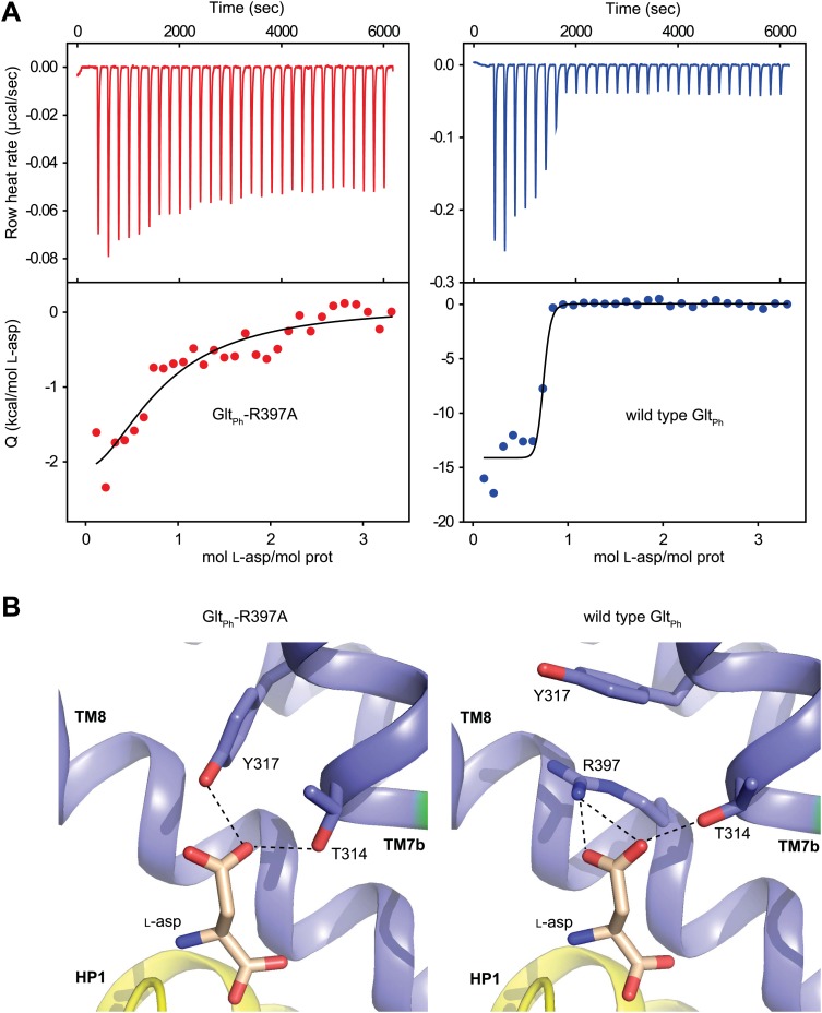
Figure 1. Substrate binding to GltPh-R397A.

(A) Raw binding heat rates measured by isothermal titration calorimetry (top) and binding isotherms (bottom) obtained for GltPh-R397A (left) and wild type GltPh (right) at 25°C in the presence of 100 mM NaCl. The solid lines through the data are fits to the independent binding sites model with the following parameters for GltPh-R397A and wild type GltPh, respectively: enthalpy change (ΔH) of −3.2 and −14.3 kcal/mol; the apparent number of binding sites (n) of 0.8 and 0.7 per monomer; dissociation constant (Kd) of 6.6 µM and 27 nM. Note that L-asp binding to the wild type transporter is too tight at 100 mM NaCl to be accurately measured in this experiment. The binding Kd has been estimated to be ∼1 nM (Boudker et al., 2007). (B) L-asp binding site in GltPh-R397A (left) and wild type GltPh (right). L-asp and residues coordinating the side chain carboxylate are shown as sticks with carbon atoms colored light brown and blue, respectively. Potential hydrogen bonds (distances less than 3.5 Å) between the L-asp side chain carboxylate and transporter residues are shown as dashed lines. Note that Y317, which forms cation-π interactions with guanidium group of R397 in wild type GltPh, interacts directly with L-asp in GltPh-R397A.

DOI: http://dx.doi.org/10.7554/eLife.02283.008

Figure 1.

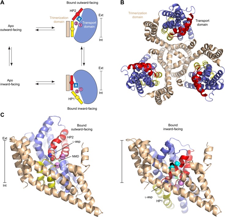
Figure 1—figure supplement 1. Alternating access mechanism in GltPh.

Figure 1—figure supplement 1.

(A) The transporter consists of a rigid trimerization domain (light brown) and a dynamic transport domain (blue, with HP1 yellow and HP2 red). The apo outward-facing transporter binds the substrate (blue square) and Na+ ions (pink circles) within the transport domain under the tip of HP2. The transport domain moves into the inward-facing state, in which the ligands are occluded from the cytoplasm by the tips of HP1 and HP2, and then released. (B) GltPh trimer viewed from the extracellular medium. (C) Single protomers of the outward-facing GltPh (PDB code 2NWX) (left), and GltPhin (PDB code 3KBC) (right). Cα atoms of the cross-linked cysteine residues and Hg2+ ion are shown as spheres. The bar next to the structures indicates approximately the thickness of the membrane, separating the extracellular (Ext) and intracellular (Int) solutions.
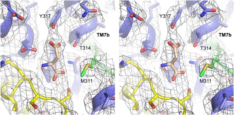
Figure 1—figure supplement 2. Structure of GltPh-R397A bound to Na+ and L-asp.

Figure 1—figure supplement 2.

Stereo view of the averaged 2Fo-Fc electron density map contoured at 1σ (grey mesh) around residues in L-asp binding site of GltPh-R397A.