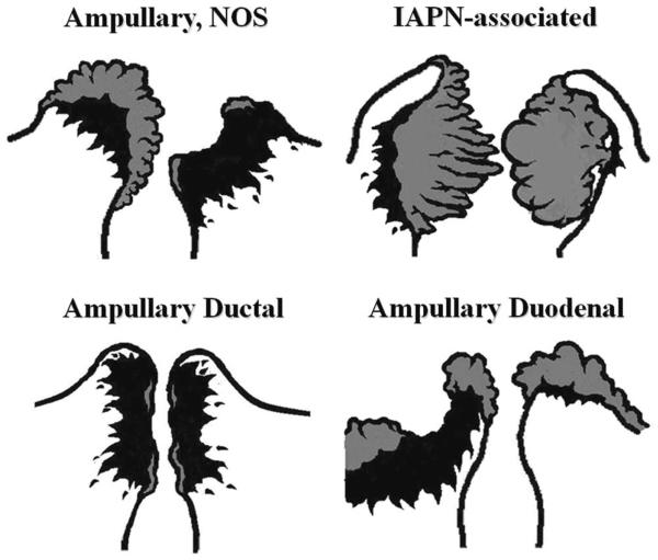
FIGURE 8.
For tumors arising in the ampullary region, careful gross examination of the distribution of the tumor and determination of the preinvasive component (adenomatous lesions that typically manifest as granular, feathery, friable, or smooth-surfaced nodular material; highlighted by gray color in this diagram) are crucial for the proper classification of the ampullary tumors. Ampullary region carcinomas comprise 4 distinct types: Intra-ampullary papillary tubular neoplasm (IAPN)-associated carcinomas are characterized by a significant preinvasive component that grows as an exophytic mass within the ampullary channel (the distal tip of the CBD and the main pancreatic duct). Carcinomas of ampullary ducts are predominantly invasive tumors that circumferentially constrict the distal end of the CBD and pancreatic duct, with minimal changes of the papilla of Vater and ampullary duodenum mucosa. Ampullary duodenal carcinomas usually arise from an adenoma of ampullary duodenum, forming bulky lesions in which the ampullary orifice is often eccentrically located. Tumors that arise from the papilla of Vater itself, as well as those not showing features that characterize the other 3 groups, are classified as ampullary carcinoma, not otherwise specified (NOS).

