Figure 1.

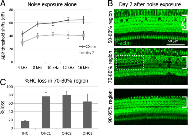
Functional and histological damage in guinea pig cochleae by noise exposure. A: ABR threshold shifts 20 min and 7 days after noise exposure in guinea pigs (n = 4). B: F-actin labeling with phalloidin in cochlear epithelia in the 50–60%, 70–80% and 90–95% regions from the cochlear apex. Brackets indicate the location of the outer hair cells. A square of dotted lines shows a patch of severe hair cell loss. C: The percent loss of hair cells in the inner hair cells (IHC), or in the first row (OHC1), second row (OHC2), or third row (OHC3) of the outer hair cells in the 70–80% region of cochlear epithelia from the apex.
