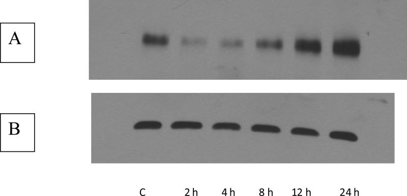
FIGURE 2.
Western blot of induction of MRP-1 protein by LPS. Total protein was isolated from RAW 264.7 cells exposed to LPS for periods ranging between 2 h and 24 h. A. Western blot using the MRPr1 antibody. The numbers below the lane indicate the time of exposure to LPS. B. Western blot using GAPDH as a loading control. The numbers below the lanes represent the time for which the cells were exposed to 100 ng/mL LPS.

