
Figure 2.
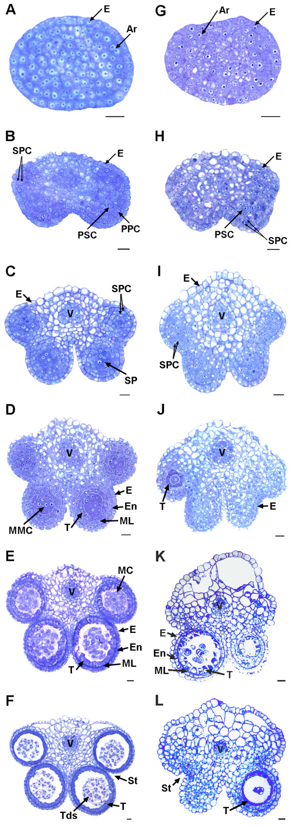
Development of fertile (A-F) and sterile (G-L) anthers in pol CMS. Bar = 10 μm for all the stages. Ar, archesporial cell; E, epidermis; En, endothecium; MC, meiotic cell; ML, middle layer; MMC, microspore mother cells; PPC, primary parietal cell; PSC, primary sporogenous cell; SP, sporogenous cell; SPC, secondary parietal cell; St, stomium; T, tapetum; Tds, tetrads; V, vascular region. A to F represent the anther development stage 2 to 7, respectively. So do G to L [25].