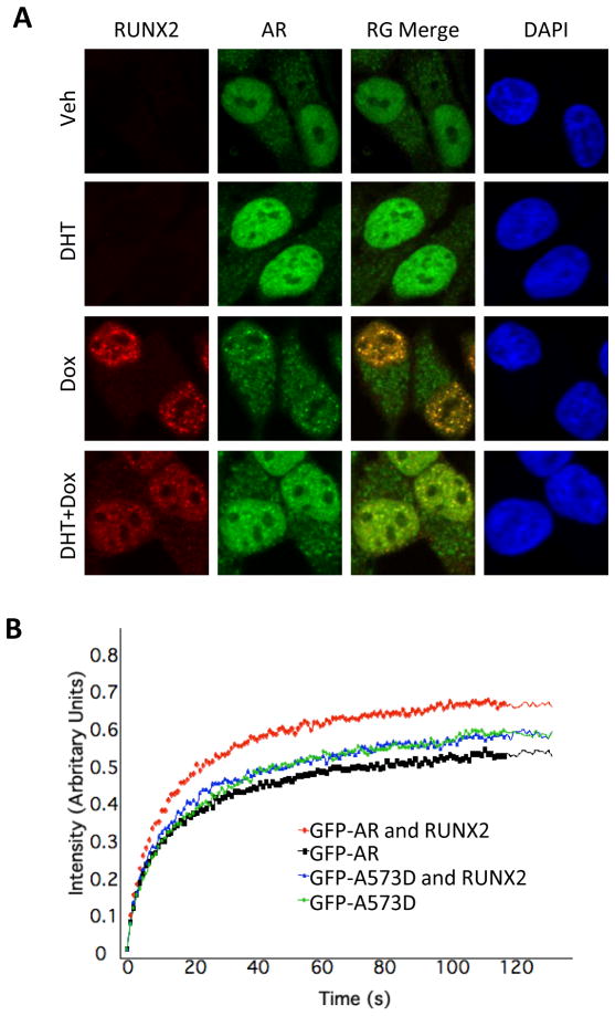
Figure 3. RUNX2 modifies AR localization and mobility in living cells.
A. C4-2B/Rx2dox cells were treated with dox and/or DHT, then immunostained and subjected to confocal microscopy to visualize the AR (green) and RUNX2 (red). DAPI (blue) demarcates the cell nucleus. B. GFP-AR or GFP-AR-A573D fusion proteins were expressed in COS7 cells either alone or together with RUNX2. The cells were treated with DHT and a portion of their nuclei was subjected to FRAP analysis. Curves represent fluorescence intensity relative to the respective pre-photobleaching levels.

